Physical Features
The shape of the egg can be described as oval, their average weight is 60 grams and its dimensions are:
|
Parameter |
Measure |
|
Weight |
60 g |
|
Volume |
53 cm3 |
|
Along circunference |
15.7 cm |
|
Widthwise circunference |
13.5 cm |
|
Egg area |
68 cm2 |
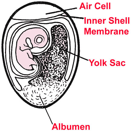
At the start of incubation, within the porous shell of the egg, three
membranes begin to develop: the amnion, chorion and allantois. The membrane
system has blood vessels that allow the developing bird to obtain oxygen and
discard carbon dioxide. Inside, there is an egg whiter (substance containing
albumin and other important components) and the yolk (which contains lots of
nutritious yolk).
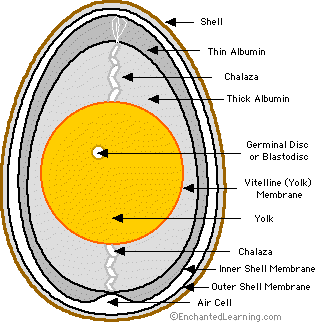
SHELL: The shell, as mentioned above, is the outer covering of the egg and is of great importance, as it acts as its physical and bacteriological barrier. It consists, for the most part, by a calcium matrix with an organic lattice, wherein the calcium is the most abundant element and of greater importance. They are also in their composition other minerals such as sodium, magnesium, zinc, manganese, iron, copper, aluminium and boron, in lower concentrations.
The shell is crossed by numerous pores that form tunnels between mineral crystals and permit gas exchange between the interior and exterior. Their number varies between 7000 and 15000. They are particularly abundant in the area of the wide pole of the egg, where the air chamber appears.
The colour of the shell, which can be white or brown depending on the breed of the hen, depends on the concentration of pigments called porphyrins, deposited in the calcium matrix and does not affect the quality or nutritional properties of the egg.
The membranes that cover the inside of the shell are two: internal and external testaceous membrane. Both surround the albumen and provide protection against bacterial penetration. The testacean membranes are strongly bonded together when the egg is laid by a hen. Shortly after laying, due to the contraction of the volume of the interior of the egg upon cooling (the body temperature of the chicken is 39 ° C, the same as layed fresh egg) air enters the thick pole, due to its bigger concentration of pores, and separate membranes in this area to form the air chamber.
The internal membrane has a fine structure of keratin fibers intertwined and the lysozyme presence in the albuminous matrix prevents entry of some microorganisms and slows the input of others. The outer membrane is much more porous and serves as a seating for the shell formation. Both membranes form around the edible part of the egg in the isthmus, which is the portion of the oviduct located between the magnum and uterus or shell gland that, as its name implies, is the place where the shell shapes.
YOLK SAC: The membrane containing the yolk or food in the yolk. It is connected to the umbilical cord and contains blood vessels. The use of the yolk is gradual at the incubation beginning, and very fast in the last five days. Initially, 25% to 30% of the yolk remains unused. Just before birth it is transferred to the body of the chick through the navel. It is absorbed during the first week of life outside the shell. Its function is nutritional. Its walls absorb albumin's food materials in the blood vessels to nourish the embryo.
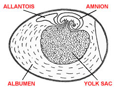
AMNION: A closed membrane sac containing amniotic fluid.
This structure develops faster than the allantois; the embryo is introduced into it. It serves to cushion the embryo against mechanical shocks, and protects against dehydration or contact with the shell. Part of this fluid is absorbed by the embryo in the later stages of development.
ALLANTOIS: It is also a membrane sac which is connected with the digestive tract, serves two functions: as a respiratory organ, bringing oxygen to the embryo and expelling carbon dioxide (gas exchange through the eggshell), and as excretory organ: the kidney excretes its products within the allantois (waste products tank that can not exit the egg).
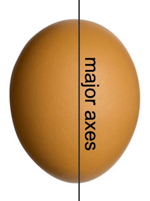
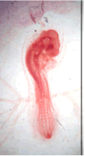
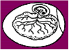
The position of the embryo is defined after 36 to 48 hours of incubation. At this time, the embryo lies in the yolk, transversely, along the minor axis.
|
|
|

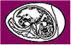
Afterwards, the head of the embryo begins to separate from the yolk and turn left. By the 5th incubation day, the embryo is near the air chamber. From the 11th day when the body of the embryo outweighs its head, it makes a left turn, which causes the body down towards the thin pole of the egg. At 14 days, the body of the embryo is located along the bigger axis of the egg, with the head facing the thick pole. This is the correct and necessary position to be taken by the chick for the birth.
|
|
|
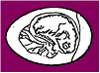
The embryo is oriented normally with his head toward the wide end of the shell. On the nineteenth day, the embryo will introduce its peak between separate membranes and will use the air chamber to breathe for the first time. The chick has the opportunity to "practice" breathing while still remaining within the shell; this allows it to undertake the final development of its various organs.
The following table shows the different signs of embryonic development during the 21 days of incubation.En la siguiente tabla se muestran los diferentes signos de desarrollo embrionario [PDF] durante los 21 días de incubación.
|
|
|
|
|
|
1 |
|

|
Appearance of veins and formation of mesodermal sac

|
|
2 |
|

|
Appearance of amniotic folds, heartbeats and blood circulation

|
|
3 |
1 cm |
|
The amnion completely surrounds the embryo, the embryo rotates anti-clockwise
|
|
4 |
1,3 cm |

|
Pigmentation of eyes; sprouts legs are longer than the wings

|
|
5 |
|
|
|
|
6 |
1,8 cm |
|
|
|
7 |
|
|
|
|
8 |
2,2 cm |
|
|
|
9 |
|
|
bird-like shape; Appearance of the mouth hole
|
|
10 |
|

|
|
|
11 |
|
|
|
|
12 |
4,5 cm |
|
|
|
13 |
|

|
|
|
14 |
|
|
|
|
15 |
|

|
|
|
16 |
|
|
|
|
17 |
|

|
|
|
18 |
|

|
|
|
19 |
|
|
|
|
20 |
|
|
The yolk sac is already in the body; the peak is
inserted into the air chamber. Pulmonary respiration and vocalization is
started.
|
|
21 |
|
|
|